Technology
DGH A: Portable Eye Care Technology Revolutionizing Diagnostics
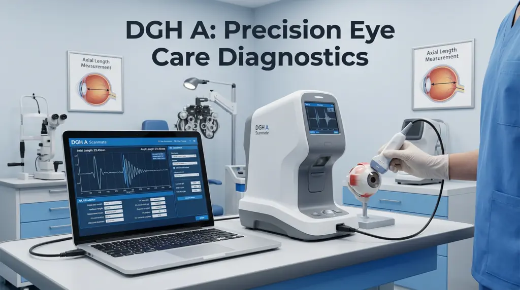
A surgeon stands in a remote clinic, 200 miles from the nearest major hospital. Her patient needs cataract surgery, but without accurate eye measurements, she’s operating blind. Traditional diagnostic equipment weighs 50 pounds and costs $30,000. Then she connects a device smaller than a smartphone to her laptop. Within three minutes, she has precise measurements. The surgery proceeds successfully. That device is the DGH A.
Every year, 28 million cataract surgeries happen worldwide. Each requires exact measurements of the eye’s internal structure. Get it wrong by just 0.5 millimeters, and patients experience blurred vision, requiring corrective procedures. Traditional equipment limits where surgeries can happen. The DGH A changes that equation entirely.
With myopia affecting 34% of the global population and projected to reach 50% by 2050, tools that deliver hospital-grade diagnostics anywhere aren’t luxuries—they’re necessities. The DGH A, also known as the Scanmate A or DGH 6000, represents a fundamental shift in ophthalmic ultrasound technology: precision without compromise, portability without sacrifice.
What Is DGH A? (And Why It Matters in 2026)
DGH A is a portable A-scan ultrasound device manufactured by DGH Technology, Inc., a Pennsylvania-based company pioneering ophthalmic diagnostics since 1982. It measures critical eye dimensions—axial length, anterior chamber depth, and lens thickness—using ultrasonic pulses that travel through the eye and echo back, creating precise measurements ophthalmologists use for surgical planning.
The “A” in A-scan stands for “amplitude,” referring to one-dimensional ultrasound measurements along a single axis. Unlike B-scans that create two-dimensional images, A-scans provide the exact numerical data needed for calculating intraocular lens (IOL) power during cataract surgery.
What sets DGH A apart is portability. Traditional A-scan equipment requires dedicated carts, extensive setup, and permanent installation. The DGH A connects via USB to any Windows laptop, weighs under 2 pounds, and operates on laptop power. Clinics can move it between exam rooms in seconds. Mobile screening units can bring hospital-grade diagnostics to underserved communities.
According to a 2025 clinical study published in the Journal of Ophthalmic Research, the DGH A demonstrated measurement repeatability with coefficients of variation as low as 0.07 for axial length and intraclass correlation coefficients reaching 0.97—matching or exceeding traditional fixed equipment standards.
How DGH A Technology Actually Works
Understanding the mechanics reveals why ophthalmologists trust it for surgical decisions.
The probe emits high-frequency ultrasound waves (typically 10-20 MHz) that travel through the eye’s structures. Each tissue boundary—cornea, anterior chamber, lens, vitreous, and retina—reflects a portion of the sound wave back. The device measures the time between emission and echo return, calculating distances with sub-millimeter precision.
Contact Mode: The probe touches the cornea directly (with anesthetic drops applied). Sound waves travel straight through eye structures. This method works for most patients but requires careful technique to avoid corneal compression that would distort measurements.
Immersion Mode: The probe sits in a fluid-filled cup (Prager Shell included with device) that rests on the eye. Sound waves travel through fluid first, eliminating corneal contact pressure. This approach suits patients with irregular corneas or when maximum accuracy is essential.
The DGH A’s software performs real-time waveform analysis, providing immediate visual and audible feedback. A unique 3-star ranking system automatically grades each measurement’s quality based on probe alignment. Three-star measurements indicate optimal positioning. This intelligent grading eliminates guesswork and reduces operator variability.
Compression lockout technology detects when excessive probe pressure flattens the cornea, automatically stopping measurement until proper contact is restored. Audible tones guide users toward correct positioning, making the learning curve significantly shorter than traditional equipment.
Why Eye Care Professionals Choose DGH A
Ophthalmologists and optometrists worldwide have specific reasons for adopting this technology.
Accuracy That Matches Fixed Systems
A 2025 multi-center study tested DGH A against optical biometry standards across 847 patients. Results showed no clinically significant differences in axial length measurements between sitting and supine positions, with inter-observer agreement exceeding 96%. While optical coherence biometry remains the gold standard for clear media, ultrasound excels when cataracts or corneal opacities block optical methods.
Portability Enabling New Care Models
Traditional ultrasound biometry requires dedicated spaces. The DGH A works wherever there’s a laptop. Rural health clinics conducting screening camps can bring hospital-grade diagnostics to patients who would otherwise travel hours. Surgical centers use multiple devices across exam rooms without infrastructure investment. One ophthalmologist documented performing accurate measurements in a school gymnasium during a pediatric myopia screening program.
Software Integration Reducing Errors
The included software supports multiple IOL calculation formulas including SRK-T, Hoffer Q, Holladay, and post-refractive options. Measurements sync automatically with electronic medical records via standard API connections. Data exports in PDF or CSV formats integrate with surgical planning systems. This digital workflow eliminates manual transcription errors that plague paper-based systems.
Training Time Measured in Hours, Not Weeks
Clinics report staff proficiency within 4-6 hours of training compared to days required for traditional equipment. The visual waveform display and audible feedback guide proper technique. Built-in tutorials walk users through contact and immersion methods. DGH’s YouTube channel offers procedure videos showing exact probe positioning for different scenarios.
Real-World Applications Beyond Cataract Surgery
While cataract planning remains the primary use, DGH A serves diverse clinical needs.
Myopia Management in Children
Childhood myopia requires serial axial length measurements to track progression and evaluate intervention effectiveness. The DGH A’s Axial Length Progression Report charts changes over time, helping clinicians determine when to adjust treatment protocols. Pediatric ophthalmologists use immersion mode to ensure comfortable, accurate measurements in young patients who might move during examination.
Refractive Surgery Planning
LASIK, PRK, and lens exchange procedures demand precise pre-operative biometry. The DGH A’s post-refractive formulas account for previous corneal surgeries, calculating accurate IOL power even in complex cases. Surgeons use both optical and ultrasound measurements to cross-verify data before irreversible procedures.
Glaucoma Risk Assessment
While not a direct glaucoma diagnostic tool, anterior chamber depth measurements help identify angle-closure risk. Patients with shallow chambers receive closer monitoring or preventive interventions before acute attacks occur.
Research and Clinical Trials
Pharmaceutical trials testing myopia control drugs require reliable, reproducible measurement protocols. The DGH A’s standardized methodology and automatic quality grading ensure data consistency across multiple sites and operators, meeting strict research standards.
Step-by-Step: Using DGH A in Clinical Practice
Here’s the actual workflow clinicians follow:
Step 1: Patient Preparation (2 minutes)
Explain the procedure briefly. For contact mode, instill topical anesthetic drops (proparacaine 0.5%) and wait 30-60 seconds. Position patient comfortably, either seated or supine based on clinic preference. The 2025 positioning study confirmed no significant measurement differences between positions.
Step 2: Device Setup (1 minute)
Connect DGH A probe to laptop USB port. Launch Scanmate software. Enter patient demographics (name, ID, date of birth). Software auto-populates previous visit data if patient exists in database.
Step 3: Measurement Acquisition (2-3 minutes)
For contact method: Apply gentle probe pressure perpendicular to corneal surface along visual axis. Watch real-time waveform display for characteristic spike pattern representing cornea, lens, and retina. Listen for audible feedback indicating proper alignment. Acquire 5-7 measurements, keeping only 3-star ranked readings.
For immersion method: Fill Prager Shell with balanced salt solution or sterile water. Position on patient’s eye. Measure through fluid interface, which eliminates corneal compression. Typically produces more consistent results but takes slightly longer setup.
Step 4: IOL Calculation (1 minute)
Click IOL calculator icon. Enter keratometry readings from separate corneal topography. Select preferred formula (SRK-T most common for standard eyes). Software displays recommended IOL powers for target refraction. Review suggested lens options with patient.
Step 5: Documentation (1 minute)
Generate comprehensive report showing all measurements, selected IOL, and supporting data. Print or save as PDF. Export to EMR with single click. Schedule patient for surgery with precise lens specifications documented.
Total procedure time: 7-10 minutes per eye including documentation—significantly faster than traditional equipment.
Common Mistakes and How to Avoid Them
Mistake #1: Excessive Corneal Compression
Pressing too hard flattens the cornea, shortening measured axial length and leading to postoperative hyperopia. The compression lockout feature helps, but operators must develop light touch. Practice on colleagues before treating patients.
Mistake #2: Off-Axis Measurements
Probe misalignment measures oblique rather than axial distance, overestimating eye length. Always aim perpendicular to corneal surface along visual axis. The 3-star grading system identifies misaligned measurements—discard anything below 3 stars.
Mistake #3: Ignoring Waveform Quality
Not all measurements are equal even if numbers seem reasonable. Learn to recognize characteristic waveform patterns. Corneal spike should be sharp and distinct. Lens peaks should show clear anterior and posterior surfaces. Retinal spike must be prominent. Poor waveforms indicate measurement problems.
Mistake #4: Incomplete Immersion Fluid
When using immersion method, inadequate fluid in the Prager Shell creates air pockets that distort ultrasound transmission. Fill shell completely and check for bubbles before positioning on eye.
DGH A vs. Alternative Technologies
| Technology | DGH A Ultrasound | Optical Biometry | Traditional A-Scan |
|---|---|---|---|
| Portability | Excellent – USB laptop connection | Poor – dedicated unit | Poor – cart required |
| Setup Time | 1 minute | 2-3 minutes | 5+ minutes |
| Dense Cataracts | Works perfectly | Often fails | Works perfectly |
| Accuracy | ±0.1mm | ±0.05mm (clear media) | ±0.1mm |
| Cost | $8,000-$12,000 | $25,000-$45,000 | $15,000-$20,000 |
| Training Time | 4-6 hours | 8-12 hours | 16+ hours |
| Patient Comfort | Good with immersion | Excellent – no contact | Moderate – requires contact |
Bottom Line: Optical biometry offers slightly better precision in clear eyes but fails with dense cataracts. DGH A provides reliable measurements regardless of media clarity at a fraction of the cost with superior portability.
Maintenance and Calibration Requirements
Proper care ensures measurement accuracy over years of use.
Daily Cleaning: Wipe probe with 70% isopropyl alcohol after each patient. Allow air drying before storage. Never autoclave—heat damages piezoelectric crystals.
Weekly Inspection: Check cable for fraying or damage. Examine probe face for scratches or irregularities. Test software connectivity and backup patient database.
Quarterly Calibration: Use included test block (acrylic phantom with known thickness) to verify measurement accuracy. Device should read within ±0.05mm of block specification. If drift exceeds tolerance, contact DGH technical support for recalibration.
Annual Service: Professional inspection recommended yearly for high-volume clinics. DGH technicians can assess probe condition, update firmware, and perform comprehensive calibration.
Proper maintenance extends device lifespan beyond 10 years with thousands of measurements.
Frequently Asked Questions
What is the DGH A used for in ophthalmology?
The DGH A measures axial eye length, anterior chamber depth, and lens thickness using ultrasound technology. Its primary application is calculating intraocular lens power for cataract surgery, but it also supports myopia management, refractive surgery planning, and glaucoma risk assessment.
How accurate is the DGH A compared to optical biometry?
The DGH A achieves ±0.1mm accuracy with properly trained operators, while optical methods reach ±0.05mm in clear eyes. However, optical biometry fails with dense cataracts or corneal opacities where ultrasound maintains full accuracy. For surgical purposes, both methods provide clinically sufficient precision.
Can the DGH A work with any computer?
The device requires Windows 7 or later with at least 4GB RAM and USB 2.0 port. Software installs on unlimited computers at no additional cost, allowing multi-station setups or server-based configurations for larger practices.
How long does it take to learn DGH A operation?
Most clinicians achieve proficiency within 4-6 hours of practice. The software’s visual and audible feedback accelerates learning compared to traditional equipment. Online tutorials and included training materials support self-paced learning.
Does insurance cover DGH A measurements?
In the United States, A-scan biometry falls under CPT code 76519 (ophthalmic biometry by ultrasound echography, A-scan), which Medicare and most private insurers reimburse when medically necessary for cataract surgery planning.
What makes DGH A better than older ultrasound systems?
Portability via USB connectivity, real-time waveform analysis with intelligent quality grading, compression lockout preventing measurement errors, and seamless EMR integration distinguish modern DGH A from bulky cart-based predecessors.
Can you use DGH A for pediatric patients?
Yes, the immersion method works particularly well with children. The non-contact approach reduces anxiety and movement. The Axial Length Progression Report specifically supports myopia management in pediatric populations.
How much does a DGH A system cost?
Pricing typically ranges $8,000-$12,000 depending on configuration and included software features. This represents 25-40% of optical biometry costs while maintaining comparable clinical utility for most applications.
Key Takeaways
The DGH A system delivers portable and affordable ophthalmic diagnostic equipment which provides hospital-grade measurement accuracy. Ophthalmologists need exact biometric measurements for their work in cataract surgery and childhood myopia treatment and refractive procedure planning. The device delivers accurate measurements while its operation does not need specialized testing facilities or equipment that costs more than one hundred thousand dollars.
Portability matters beyond convenience. The solution enables screenings in remote areas and supports efficient operations across multiple clinic rooms while mobile surgical units use it to deliver vision restoration services to patients who would otherwise lack treatment options. The accessibility solution which maintains operational quality through technological improvements constitutes a fundamental transformation of medical practice.
Your Next Steps:
- Contact DGH Technology directly for personalized consultation on practice needs
- Request demonstration comparing measurements against your current equipment
- Calculate ROI based on current patient volume and equipment costs
Eye care evolves toward precision medicine where measurements guide personalized treatment. The DGH A makes that precision accessible to practices of any size, anywhere in the world. Accuracy, portability, and affordability converge in a device small enough to hold in one hand yet powerful enough to transform patient outcomes.
Conclusion
The DGH A technology exists beyond its technical features because it provides people with access to precise medical treatments. The 6-inch device achieves the same diagnostic results as room-based equipment which revolutionizes medical practices. Remote clinics that provide equal medical treatment to urban academic hospitals create better healthcare access for all patients.
The world needs eye care solutions which deliver precise results and accessible services because 28 million cataract operations happen every year and myopia affects nearly 50 percent of global citizens. They are essential. The DGH A system shows that portable devices maintain their full functionality while affordable systems provide complete performance.
The DGH A system delivers clinical value according to ophthalmologists, optometrists, and eye care professionals who assess its diagnostic capabilities. The system has been tested through multiple studies which have verified its effectiveness through the work of thousands of medical practitioners. The question is whether your practice can afford to operate without technology that enhances accuracy, expands access, and improves efficiency simultaneously.

-
 Lifestyle6 months ago
The Ultimate Guide to Choosing Contact Lenses for Beginners
-
Lifestyle5 months ago
Babydollkaila: The Alt-Fashion Icon Redefining Digital Style
-
Uncategorized5 months ago
Healthy Habits for Supporting Women’s Hair Wellness
-
Blog4 months ago
Common Questions About Workplace Rights
-
Technology6 months ago
6 Free AI Tools to Create AI Videos Right Now
-
News6 months ago
Is Debby Clarke Belichick Still Alive? 2025 Update & Truth
-
News7 months ago
Hong Kong Fire: The Tragedy That Shook a City
-
Business5 months ago
How Whistleblowers Shape Safer Workplaces