Picture yourself sitting in a doctor’s office, worried about persistent headaches that won’t go away. Your physician orders an MRI scan, and within hours, a detailed diag image reveals exactly what’s happening inside your brain—no surgery required, no guesswork, just clear answers.
That’s the power of modern diagnostic imaging, and it’s saving lives every single day.
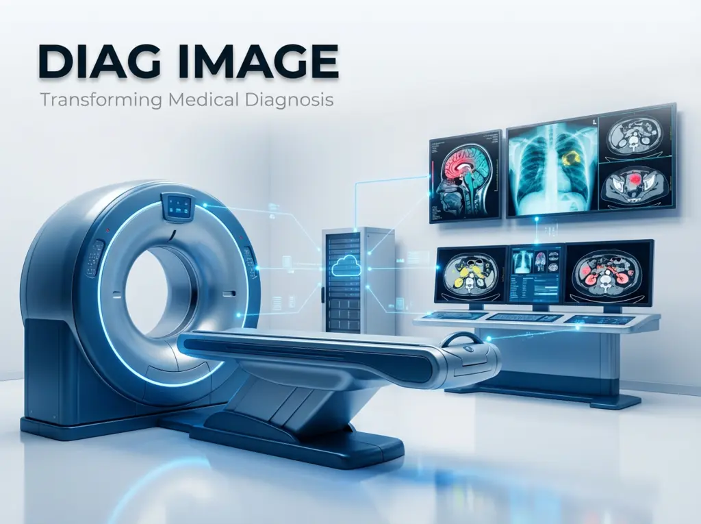
Medical imaging has evolved from grainy X-rays to crystal-clear 3D reconstructions that let doctors see inside your body with stunning precision. Whether you’re dealing with a sports injury, monitoring cancer treatment, or checking on a developing baby, diag image technology plays a crucial role in your healthcare journey.
What Exactly Is a Diag Image?
A diag image, the abbreviation for diagnostic image, represents a visual image of the internal structure of your body made with the help of a special medical device. The images are of great assistance to the medical practitioners in diagnosing the diseases, deciding on the treatments, and checking how well the therapies are working.
Just imagine, giving your doctor superpower X-ray vision, but with options even better than those of Superman. Today’s imaging methods depict everything: from the hardest bones to the softest tissues, from blood flow to the activity of metabolism.
The method operates by applying different types of energy—electromagnetic radiation, sound waves, magnetic fields, or radioactive tracers—to your body and subsequently obtaining very clear images of the inside.
Types of Diagnostic Imaging You Might Encounter
X-Rays: The Classic Standard
X-rays remain the most common type of diagnostic imaging. These electromagnetic rays pass through your body, creating images that highlight bones and dense tissues.
Your dentist uses them to spot cavities. Emergency rooms rely on them to identify fractures. They’re quick, affordable, and remarkably effective for many common conditions.
CT Scans: Layered Precision
Computed Tomography (CT) scans combine multiple X-ray images taken from different angles to create cross-sectional “slices” of your body. Radiologists can stack these slices to build 3D models, revealing injuries or diseases that standard X-rays might miss.
CT scans excel at detecting internal bleeding, diagnosing cancer, and evaluating complex fractures. The entire scan typically takes less than 10 minutes.
MRI: Soft Tissue Specialist
Magnetic Resonance Imaging uses powerful magnets and radio waves to generate incredibly detailed images of organs, soft tissues, and the nervous system. Unlike X-rays and CT scans, MRIs don’t use ionizing radiation.
Athletes often get MRIs to diagnose torn ligaments or cartilage damage. Neurologists use them to detect brain tumors, strokes, and multiple sclerosis. The trade-off? MRI scans take longer—usually 30 to 60 minutes—and the machine can feel confining.
Ultrasound: Sound Wave Imaging
Ultrasound technology bounces high-frequency sound waves off internal structures, creating real-time moving images. You’ve probably seen ultrasound used during pregnancy to check on a developing baby.
Beyond obstetrics, ultrasound helps diagnose heart problems, examine abdominal organs, and guide needle biopsies. It’s completely non-invasive and doesn’t involve radiation exposure.
PET Scans: Metabolic Mapping
Positron Emission Tomography (PET) scans inject a small amount of radioactive tracer into your bloodstream, then track where it accumulates in your body. Cancer cells, which consume more glucose than normal cells, light up brightly on PET scans.
Oncologists combine PET with CT imaging to locate tumors, determine cancer staging, and evaluate treatment effectiveness.
Why Diagnostic Imaging Matters More Than Ever
About 12 million Americans receive incorrect or delayed diagnoses each year, according to healthcare research. Image diagnosis technology helps reduce these errors by providing objective visual evidence that supplements clinical examination.
Speed Saves Lives
When someone arrives at the emergency room with stroke symptoms, every minute counts. Brain cells die rapidly without adequate blood flow. A CT scan can identify whether the stroke resulted from a blood clot or bleeding in just minutes, completely changing the treatment approach.
That speed difference can mean the gap between full recovery and permanent disability.
Catching Problems Early
Regular mammograms detect breast cancer at stages when tumors are too small to feel during physical exams. Early detection dramatically improves survival rates—the five-year survival rate for localized breast cancer exceeds 99%.
Similar stories play out across medicine. Low-dose CT scans find lung cancer in high-risk patients. Ultrasounds catch abdominal aneurysms before they rupture. Diagnostic imaging turns potential emergencies into manageable health conditions.
Avoiding Unnecessary Surgery
Before modern imaging existed, doctors sometimes performed exploratory surgery just to figure out what was wrong. Today, a diag image provides that same insight without making a single incision.
This means less risk, shorter recovery times, and lower healthcare costs.
How AI Is Changing Diagnostic Imaging
Artificial intelligence is revolutionizing how radiologists interpret medical images. AI algorithms can analyze thousands of scans to identify patterns that might escape human eyes, flagging suspicious areas for closer examination.
A 2020 study in The Lancet Digital Health found that AI systems matched or exceeded radiologist performance in detecting breast cancer from mammograms. The technology doesn’t replace doctors—it enhances their capabilities, reducing interpretation errors by up to 30% for specific imaging tasks.
Some AI systems now detect lung nodules, identify bone fractures, measure tumor sizes, and even predict which lesions are likely to be cancerous. Radiologists review the AI findings and make final diagnostic decisions, combining machine precision with human judgment.
What Happens at a Diagnostic Imaging Center
If you’ve never visited a dedicated imaging center, here’s what you can typically expect:
You’ll check in and complete paperwork about your medical history, current symptoms, and any allergies. A technician explains the procedure and answers your questions. Depending on the exam type, you might need to change into a gown or remove jewelry.
During the scan, you’ll usually lie on a table that slides into or past the imaging equipment. For most scans, you need to stay very still—even slight movement can blur the images. Some procedures require you to hold your breath briefly.
The actual imaging often takes just minutes, though MRI scans run longer. Afterward, a radiologist reviews your diag images and sends a detailed report to your referring physician, usually within 24 to 48 hours.
Many centers now provide patient portals where you can view your images and reports online, giving you more direct access to your health information.
Common Uses Across Medical Specialties
Emergency Medicine: Quickly diagnoses traumatic injuries, internal bleeding, and acute conditions like appendicitis or kidney stones.
Cardiology: Evaluates heart structure and function, detects blockages in coronary arteries, and monitors pacemaker placement.
Oncology: Locates tumors, determines cancer spread, plans radiation therapy, and assesses treatment response.
Orthopedics: Examines bone fractures, joint problems, spinal conditions, and sports injuries with precise measurements for surgical planning.
Neurology: Detects brain tumors, evaluates stroke damage, diagnoses multiple sclerosis, and monitors degenerative diseases.
Obstetrics: Monitors fetal development, estimates due dates, detects birth defects, and guides procedures during pregnancy.
Understanding the Risks and Limitations
While diagnostic imaging provides tremendous benefits, you should understand potential drawbacks:
Radiation Exposure: X-rays, CT scans, and PET scans use ionizing radiation. A single scan delivers minimal exposure, but repeated imaging over time increases cumulative risk. Your doctor weighs these risks against diagnostic benefits.
Cost Considerations: Advanced imaging can be expensive. A basic X-ray might cost $100, while a PET-CT scan can run $3,000 or more. Insurance coverage varies, and you may need pre-authorization for certain exams.
False Positives: Sometimes imaging detects abnormalities that turn out to be harmless, leading to unnecessary biopsies or additional testing that causes anxiety.
Contrast Reactions: Some procedures use contrast dyes to improve image quality. These dyes occasionally cause allergic reactions, though serious complications are rare.
Making the Most of Your Imaging Experience
If your doctor orders diagnostic imaging, you can take steps to ensure the best possible results:
Ask questions about why you need the test, what it will show, and how results will guide your treatment. Understanding the purpose helps reduce anxiety.
Follow preparation instructions carefully. Some scans require fasting, stopping certain medications, or drinking contrast material beforehand.
Mention claustrophobia if you feel anxious about enclosed spaces. Technicians can often provide strategies to help you stay comfortable during MRI scans.
Request copies of your images and reports for your personal records, especially if you see multiple doctors or might need second opinions.
The Future of Diagnostic Imaging
The medical imaging discipline endures a constant change owing to the technological innovations and advancements. Ultrasound machines that are portable are so small that they can fit into the pockets of the doctors; so, the diagnostic power is being carried to the most remote medical institutions and emergency situations. Imaging data is transformed into 3D printed physical models that can be viewed and touched by the surgeons before carrying out the complicated operations.
Molecular imaging techniques are able to visualize the biological activities at the level of individual cells, and consequently, they can detect the diseases many years earlier than the current techniques. In addition, the systems that are under development have the potential to integrate multiple imaging modalities in one go thus providing exceptionally detailed diagnostic information from a single scan.
On the other hand, tech advancement means that the operators will have to deal with fast scans, low ionizing radiation, high-quality images, and AI support that will ensure interpretation not just precise but also broadly available.
Taking Action: When to Request Imaging
Imaging tests cannot be ordered by yourself — doctors decide these things taking into account your clinical signs, medical history, and physical examination findings. Nonetheless, you can still request appropriate testing if the doubts persist.
Talk to your doctor about imaging when the treatment isn’t helping and the symptoms have worsened, or if new problems appeared or you’re still not getting clear answers about what is wrong. Ask precisely: “Could imaging assist in ruling out serious causes?” or “Would a scan give information that is useful for my treatment plan?”
Keep in mind that an MRI is not needed for every pain. Your doctor weighs the diagnostic advantages against the costs and risks and orders tests only when the results will make a significant difference to your treatment.
Frequently Asked Questions
How long does it take to get diag image results?
Most imaging centers send reports to your doctor within 24 to 48 hours. Emergency scans receive immediate interpretation. Complex cases might take longer if radiologists need to consult specialists.
Are diagnostic images painful?
The imaging process itself doesn’t hurt. You might feel mild discomfort from lying still on a hard table or holding uncomfortable positions. Some contrast injections cause brief warmth or unusual taste sensations.
Can I eat before getting imaging done?
This depends on the specific test. X-rays and most MRIs don’t require fasting. CT scans with contrast usually require you to avoid food for several hours beforehand. Always follow the preparation instructions you receive.
How much radiation am I exposed to during imaging?
A chest X-ray delivers about 0.1 millisieverts (mSv) of radiation—roughly equivalent to 10 days of natural background radiation. CT scans vary from 2 to 20 mSv depending on the body area scanned. Your doctor considers these doses when ordering tests.
Will my insurance cover diagnostic imaging?
Most health insurance plans cover medically necessary imaging when your doctor orders it. You may need pre-authorization for expensive tests like MRI or PET scans. Check your specific plan details and get approval before scheduling when required.
Can I request a specific type of imaging?
Your doctor determines which imaging type best answers the clinical question. Different technologies excel at visualizing different tissues and conditions. Trust your physician’s expertise while asking questions about the recommendation.
How accurate are diag images?
Accuracy varies by condition, imaging type, and the radiologist’s skill. No test is perfect—some cancers don’t show up on initial scans, and some benign findings look suspicious. Your doctor combines imaging results with other clinical information for the most accurate diagnosis.
Final Thoughts
The use of diagnostic imaging has undergone a significant change, and it is now considered an essential part of healthcare, saving countless lives in the process. The visual documentation that diag image technology provides, is needed by doctors to perform your care in the most efficient way, whether you are having a routine screening, looking into mysterious symptoms, or checking your chronic disease.
The next time you come across an X-ray, CT scan, or MRI machine, just think that you are witnessing the art of human body exploration with a technology that was considered magical only a few decades ago. These tools not only help in diagnosing the diseases in an early stage but also prevent the matter of surgery and deliver quicker and accurate diagnosis.
Being up to date with your health, always asking questions during imaging, and having confidence in these robust diagnostic instruments working towards your wellness and imaging are the keys.